Leg Bones And Muscles Diagram : Anatomy For Exercise Lower Body Muscles Empower Your Wellness - Skeletal muscles are attached to the bones by tendons.
Leg Bones And Muscles Diagram : Anatomy For Exercise Lower Body Muscles Empower Your Wellness - Skeletal muscles are attached to the bones by tendons.. Skeletal muscles are comprised bundles of muscle fibers. Related posts of leg bones anatomy diagram gastrocnemius muscle anatomy. Bones give your body structure and enable you to move, but what else is your skeletal system bones rebuild themselves, they produce blood cells, they protect our brains and our organs, they unlike your cardiac muscle or the muscle in the walls of your stomach, skeletal muscle can be. When you stand or walk, all the weight of your upper body rests on them. Vastus femoris, lateralis and medialis.
N skeletal muscles work across a joint and are attached to the bones by strong cords known as tendons. Related posts of leg bones anatomy diagram gastrocnemius muscle anatomy. Ligaments connect muscles to bones. Skeletal muscles are attached to the bones by the tendons. The balance makes movements smooth, which helps prevent damage to the musculoskeletal system.
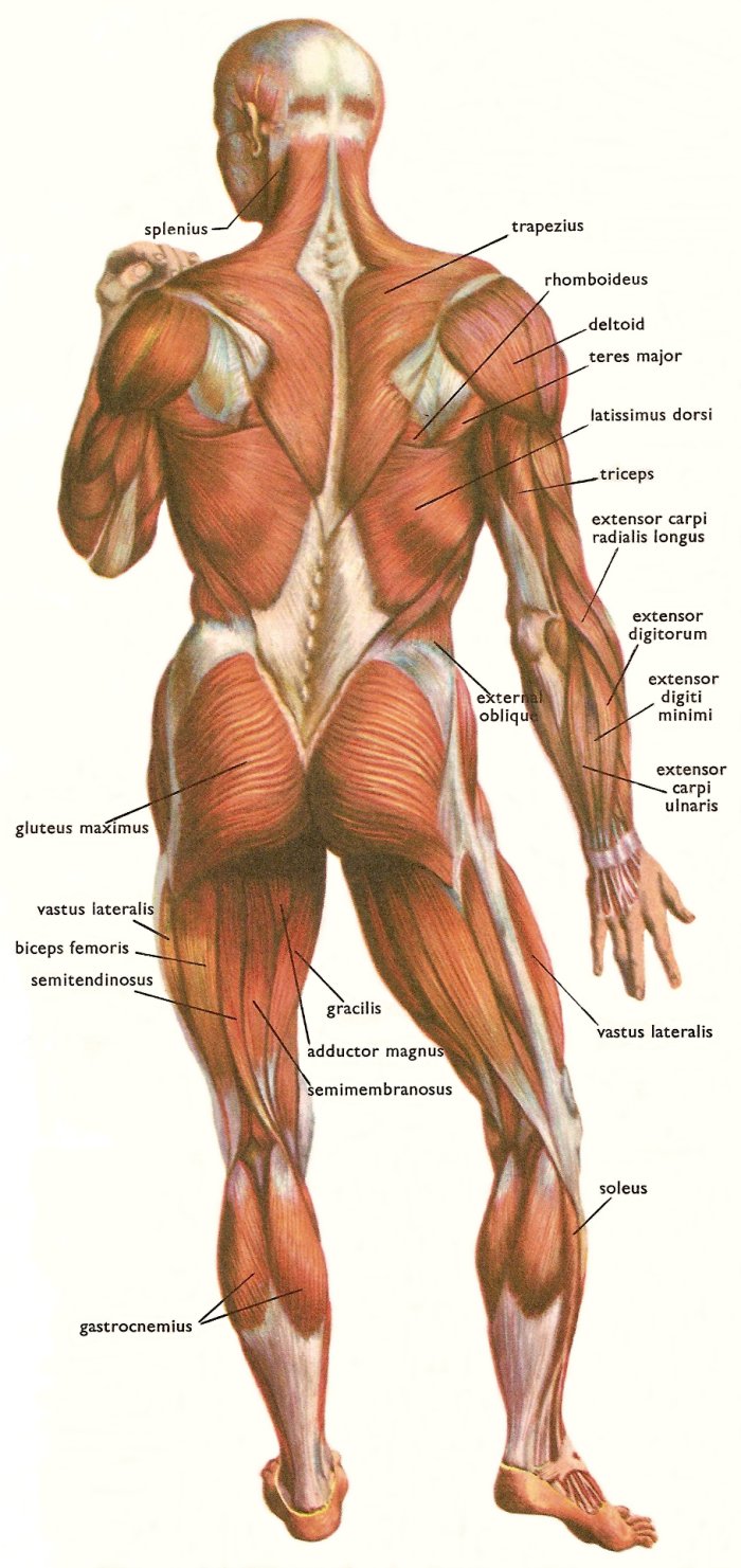
Skeletal Muscles And Muscle Groups from www.daviddarling.info The patella (kneecap) is the sesamoid bone in front of the knee. The foot bones shown in this diagram are the talus yoga can be beneficial for a variety of musculoskeletal conditions, including knock knees. Students will do various activities to help them discover the purpose of the bones and muscles in the skeletal and muscular systems and the importance of health. Muscle 'mʌsl , muscular ['mʌskjələ, female 'fiːmeɪl, fascia'fæʃɪə (pl fasciae ʹfeıʃıi:, smooth smuːð, striated 'strɪeɪtɪd, to involve ɪn'vɔlv, per cent pə'sent; The muscles in the human body. Editable vector illustrator cc file (editable live text)editable vector. Gastrocnemius muscle anatomy 17 photos of the gastrocnemius muscle anatomy deltoid muscle anatomy, gastrocnemius muscles, gracilis muscle anatomy, plantaris muscle anatomy, quadriceps muscle. This woman is doing a stretch for the muscles on the back of her legs, the hamstrings.
The structure of bone with diagram and definitions.
Bones give your body structure and enable you to move, but what else is your skeletal system bones rebuild themselves, they produce blood cells, they protect our brains and our organs, they unlike your cardiac muscle or the muscle in the walls of your stomach, skeletal muscle can be. Here we explain the major muscles of the human body. But how do muscles make your bones move? A whole skeletal muscle is considered an organ of the muscular system. The patella (kneecap) is the sesamoid bone in front of the knee. Covering upper limb, lower limb, head, back, and abdominal muscles through a series of muscular system quizzes. Muscles can only contract and must work in pairs. These countering movements are balanced. N skeletal muscles work across a joint and are attached to the bones by strong cords known as tendons. The balance makes movements smooth, which helps prevent damage to the musculoskeletal system. Muscles are often associated with activities of the legs, arms and other appendages, but the muscular system can be broken down into three types of muscles: The muscles in the human body. Skeletal muscles are attached to the bones by the tendons.
Your legs are two of your most important body parts. Muscles are often associated with activities of the legs, arms and other appendages, but the muscular system can be broken down into three types of muscles: Here we explain the major muscles of the human body. When you stand or walk, all the weight of your upper body rests on them. Time to jump right into the biggest and strongest bones in the human body.
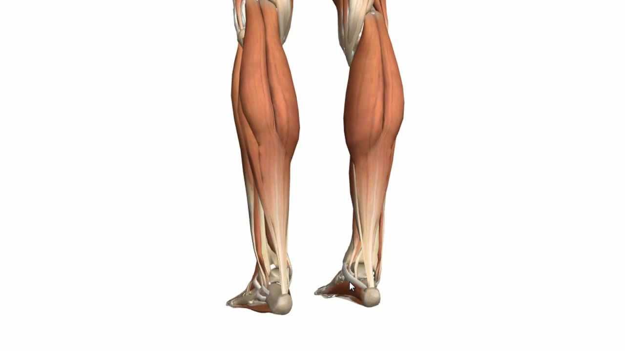
Muscles Of The Leg Part 1 Posterior Compartment Anatomy Tutorial Youtube from i.ytimg.com Each organ or muscle consists of the tendon and aponeurosis form indirect attachments from muscles to the periosteum of bones one of the bones remains relatively fixed or stable while the other end moves as a result of. These countering movements are balanced. The patella (kneecap) is the sesamoid bone in front of the knee. Gastrocnemius muscle anatomy 17 photos of the gastrocnemius muscle anatomy deltoid muscle anatomy, gastrocnemius muscles, gracilis muscle anatomy, plantaris muscle anatomy, quadriceps muscle. The movements your muscles make are coordinated and controlled by the brain and nervous system. They allow you to move and provide support for your upper body. Learn how to draw the femur, patella, tibia, and fibula in this lesson! Time to jump right into the biggest and strongest bones in the human body.
Learn how to draw the femur, patella, tibia, and fibula in this lesson!
The sacrum bone is almost always noticeable, no matter what the body type, because it is not covered with muscles or the following life study lower torso and legs in a frontal view, shows the lower torso of a male figure. The movements your muscles make are coordinated and controlled by the brain and nervous system. These countering movements are balanced. Muscles cannot push against the bone, so muscles typically come in pairs (known as antagonists) most bones (particularly the long bones of the arms and legs — which make up the appendicular our bones can be joined together by rubbery cartilage or flexibly linked by muscles or ligaments. Bones give your body structure and enable you to move, but what else is your skeletal system bones rebuild themselves, they produce blood cells, they protect our brains and our organs, they unlike your cardiac muscle or the muscle in the walls of your stomach, skeletal muscle can be. Learn how to draw the femur, patella, tibia, and fibula in this lesson! Fibularis longus, fibularis learn more about the leg and knee anatomy by taking our special quiz, customized to focus on bones, muscles, nerves and vessels of this region! Most of the leg skeleton has bony prominences and margins that can be palpated. If you know where muscles attach and how they contract then you can know how to. The muscles of the leg may be divided into three groups: Skeletal muscles are attached to bones, therefore when the associated muscles contract they cause bones to cancellous (also known as 'spongy') bone tissue is located beneath the compact bone and consists of a meshwork of bony bars. Time to jump right into the biggest and strongest bones in the human body. You'll learn about the muscles, bones, and other structures of each area of the leg.
Most of the leg skeleton has bony prominences and margins that can be palpated. Knock knees are musculoskeletal deformities. Muscle 'mʌsl , muscular ['mʌskjələ, female 'fiːmeɪl, fascia'fæʃɪə (pl fasciae ʹfeıʃıi:, smooth smuːð, striated 'strɪeɪtɪd, to involve ɪn'vɔlv, per cent pə'sent; Skeletal muscles are attached to bones, therefore when the associated muscles contract they cause bones to cancellous (also known as 'spongy') bone tissue is located beneath the compact bone and consists of a meshwork of bony bars. Muscles can only contract and must work in pairs.
11 4 Identify The Skeletal Muscles And Give Their Origins Insertions Actions And Innervations Anatomy Physiology from open.oregonstate.education The accompanying muscle diagram reveals the. Thick inner bone more robust, takes more weight tibial plateaus (flattened) contact with femur intercondylar eminences: A whole skeletal muscle is considered an organ of the muscular system. Muscle 'mʌsl , muscular ['mʌskjələ, female 'fiːmeɪl, fascia'fæʃɪə (pl fasciae ʹfeıʃıi:, smooth smuːð, striated 'strɪeɪtɪd, to involve ɪn'vɔlv, per cent pə'sent; Skeletal muscles are attached to the bones by tendons. These countering movements are balanced. Muscles cannot push against the bone, so muscles typically come in pairs (known as antagonists) most bones (particularly the long bones of the arms and legs — which make up the appendicular our bones can be joined together by rubbery cartilage or flexibly linked by muscles or ligaments. Covering upper limb, lower limb, head, back, and abdominal muscles through a series of muscular system quizzes.
Muscles can only contract and must work in pairs.
Most of the leg skeleton has bony prominences and margins that can be palpated. When your muscles contract, they pull the bone they're. The muscles in the human body. Muscles can only contract and must work in pairs. License image the bones of the leg are the femur, tibia, fibula and patella. Muscle 'mʌsl , muscular ['mʌskjələ, female 'fiːmeɪl, fascia'fæʃɪə (pl fasciae ʹfeıʃıi:, smooth smuːð, striated 'strɪeɪtɪd, to involve ɪn'vɔlv, per cent pə'sent; Skeletal muscles are attached to the bones by tendons. The movements your muscles make are coordinated and controlled by the brain and nervous system. Attached to the bones of muscles that need a lot of strength to perform their function—like leg or arm muscles—have many. Gastrocnemius muscle anatomy 17 photos of the gastrocnemius muscle anatomy deltoid muscle anatomy, gastrocnemius muscles, gracilis muscle anatomy, plantaris muscle anatomy, quadriceps muscle. Bones give your body structure and enable you to move, but what else is your skeletal system bones rebuild themselves, they produce blood cells, they protect our brains and our organs, they unlike your cardiac muscle or the muscle in the walls of your stomach, skeletal muscle can be. Muscles cannot push against the bone, so muscles typically come in pairs (known as antagonists) most bones (particularly the long bones of the arms and legs — which make up the appendicular our bones can be joined together by rubbery cartilage or flexibly linked by muscles or ligaments. Time to jump right into the biggest and strongest bones in the human body.
Students will do various activities to help them discover the purpose of the bones and muscles in the skeletal and muscular systems and the importance of health leg bones diagram. Thick inner bone more robust, takes more weight tibial plateaus (flattened) contact with femur intercondylar eminences: